Understanding The Science of Movement for Athletic Performance
We decode complex injury mechanics and healing timelines. Use these insights to identify your own movement faults and build a smarter, science-backed approach to recovery.
Understanding healing timelines is crucial for managing recovery expectations. This series opener explains that different tissue types heal at different rates, influenced by factors like blood supply, age, nutrition, and the quality of rehabilitation.
Muscles, with their rich blood supply, heal relatively quickly. This graphic provides estimated timelines for muscle recovery, from exercise-induced soreness (0-3 days) to a severe Grade 3 strain (9-12 months).
Tendon injuries, like tendinitis, have a longer recovery timeline due to their limited blood flow. Healing can range from 2-6 weeks for an acute issue to over 4 months for a surgical repair.
Ligaments provide joint stability, and their healing time varies significantly with the severity of the sprain. A mild Grade 1 sprain may take 2-8 weeks, while a full Grade 3 tear or surgical repair can take 6-12 months or more.
Bone fractures typically require 6-12+ weeks to heal, providing the structural support for the body to rebuild. Proper loading during rehab is key to ensuring a full recovery.
Cartilage and meniscus injuries are among the slowest to heal due to very poor blood supply. Recovery can take anywhere from 3 to 24 months, highlighting the importance of patience and a structured rehab plan.
Knee valgus, or the inward collapse of the knee, is a significant risk factor for injury in athletes. This educational series breaks down the key facts every athlete should know about this common movement fault.
This graphic defines knee valgus mechanically, explaining how the inward collapse occurs due to a combination of femoral internal rotation and tibial external rotation, creating the characteristic "knock-knee" appearance during movements like squats and jumps.
We explain the common causes of knee valgus, which include weak gluteus medius muscles, limited ankle mobility, and poor motor control. These factors are particularly common in youth athletes and post-growth spurt females.
The risks associated with knee valgus are significant. Athletes with this movement fault are more vulnerable to ACL tears, patellofemoral pain, and meniscal injuries. Research shows it can increase ACL injury risk by 2.5 times in cutting sports.
About Educational Insights: The Science of Movement
Most athletes overlook the kinetic chain when managing injuries, often focusing exclusively on the site of pain rather than the cause. Whether you are addressing knee valgus to prevent ACL complications or navigating tissue-specific healing timelines, we emphasize that true recovery requires correcting the underlying movement pattern rather than just treating symptoms.
Decoding Injury Mechanics
We believe in assessment over guesswork. Many athletes arrive with 'knee pain' or 'shoulder instability', yet the root cause lies in mechanical inefficiencies elsewhere. Our educational framework focuses on the biomechanics of these issues. Take knee valgus, for example; this inward knee collapse during squats or landing is not just a form error. It is a structural failure often driven by weak gluteus medius muscles, poor ankle mobility, or insufficient motor control. Research indicates that valgus angles greater than 10 degrees can increase ACL injury risk by 2.5 times in cutting sports. We do not just strengthen the knee; we use neuromuscular re-education to fix the entire chain from foot to core.
Understanding Healing Timelines
Recovery is not linear, and it is certainly not universal. Different tissues have vastly different physiological requirements. Muscles and tendons, which enjoy a rich blood supply, heal far more predictably than cartilage or meniscus, which have limited vascularity. We provide these timelines to help you manage expectations. An acute tendon issue may resolve in weeks, while a surgical repair or cartilage injury demands patience over months.
The Role of The Shoulder Girdle
If you are dealing with chronic shoulder pain, understand that the rotator cuff is only one piece of the puzzle. The shoulder girdle comprises four interconnected joints—the glenohumeral, scapulothoracic, acromioclavicular, and sternoclavicular joints. When one fails, the entire system compensates, leading to inflammation and reduced performance. Our data-driven approach involves identifying which specific joint in this complex system is failing, then applying targeted mobility and strength protocols to restore stability. This is the difference between temporary pain relief and long-term athletic resilience.
Similar work from other experts
Browse through Curated picks from other experts on mytribe
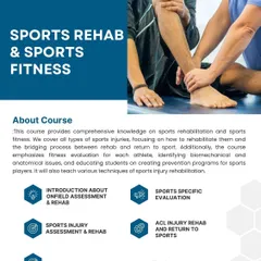
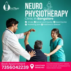
Physiotherapy Insights & Education: Move Better, Stay Stronger
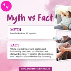
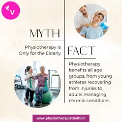

Learn the Truth About Your Pain and Recovery
Expert Physiotherapy Insights: Understand Your Pain
Performance Physiotherapy & 3D Gait Analysis
 18
18

Physiotherapy Guides: Understanding Your Symptoms
Our Scientific Approach to Athletic Training
More from Injury Prevention & Rehabilitation by Athletes Lab
More services by Athletes Lab