Anatomy and Injury Guides: Understanding Your Pain
Don’t guess what’s wrong with your knee or ankle. Browse our visual guides to understand your injury, then let us help you move better.
This video shows a clear example of patellofemoral joint hypermobility, where the kneecap moves excessively. This condition is common in athletes and can cause pain and instability, but it can be managed effectively with targeted strengthening exercises.
Turf toe is a common injury in sports, caused by hyperextension of the big toe joint. This diagram explains the mechanism of injury, which damages the ligaments and can cause significant pain during push-off movements like running or jumping.
This illustration shows the difference between normal knee alignment, bowlegs, and knock knees. Misalignment can put abnormal stress on your joints and lead to pain over time. We can help correct these issues with personalized exercise programs.
Ankle sprains are one of the most frequent sports injuries. This guide shows the different ligaments that can be affected and outlines a comprehensive treatment approach, including mobility exercises, strengthening, and taping to ensure a full recovery.
Can you identify the fracture in this X-ray? This is a patellar, or kneecap, fracture. Understanding medical imaging is part of our diagnostic process to create the most effective and safe rehabilitation plan for our patients.
The pectoral girdle muscles, including the rhomboids and serratus anterior, are essential for shoulder stability and upper body movement. This anatomical chart explains their function and why strengthening them is key to good posture and injury prevention.
This 3D animation shows the complex muscle framework of the upper body. Understanding how muscles like the pectoralis major and deltoids work together during exercises like the bench press is crucial for proper form and preventing injury.
The deep back muscles are the foundation of a strong, stable spine. This animation provides a detailed look at these crucial muscles, which are essential for maintaining posture and preventing common issues like lower back pain.
This video highlights the latissimus dorsi, or "lats," one of the largest muscles in the back. It demonstrates their primary role in exercises like the lat pull-down, which is key for building back strength and a V-taper physique.
Wrist fractures can occur in several ways. This diagram illustrates common types like the Colles and Smith fractures, showing how the angle of the break differs. Proper diagnosis is critical for a successful recovery and regaining full hand function.
About Anatomy & Injury Guides
When you come to our clinic with an injury, we don't just look at the spot where it hurts. We look at the mechanics. For example, a runner with persistent knee pain often has a 'stiff big toe' (hallux limitus) forcing their gait to compensate. By understanding how your body’s levers work together, we create a recovery plan that fixes the root cause, not just the symptom.
Why do we show you the anatomy? Because knowing how your body functions is the first step toward getting it back to peak performance. We often see patients who have been dealing with 'nagging' issues—like Patellofemoral joint hypermobility or Intersection Syndrome—without knowing why they happen.
The 'Root Cause' Approach
Most pain isn't just about the area where you feel the discomfort. That ankle sprain might stem from weakness in your hip stabilizer muscles. That wrist pain from weightlifting might be simple biomechanics gone wrong. Our guides help you spot these patterns early, before they turn into chronic issues.
How We Use This Knowledge
Whether it is correcting knock-knees or managing turf toe, we combine these visual insights with hands-on modalities like IASTM, cupping, and dry needling. You aren't just a patient with a 'bad back'; you are an athlete, a professional, or a grandparent who needs to walk without stiffness. Our goal is to translate complex anatomy into a clear plan for your body.
What to Expect
If you recognize these patterns in yourself, don't wait for them to worsen. Bring your symptoms to our clinic in Noida, Indirapuram, or Pandav Nagar. We will map out your movement patterns, identify the trigger, and build a program that brings you back to your best.
Similar work from other experts
Browse through Curated picks from other experts on mytribe
 18
18

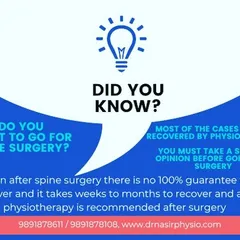
Physiotherapy Guides: Understanding Your Symptoms
 27
27
Understanding The Science of Movement for Athletic Performance
 13
13
Understanding Your Pain: Identifying Root Causes
 20
20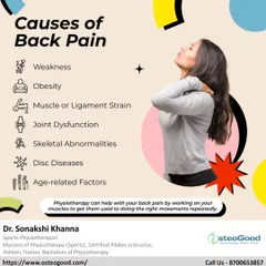


Your Guide to a Pain-Free Life
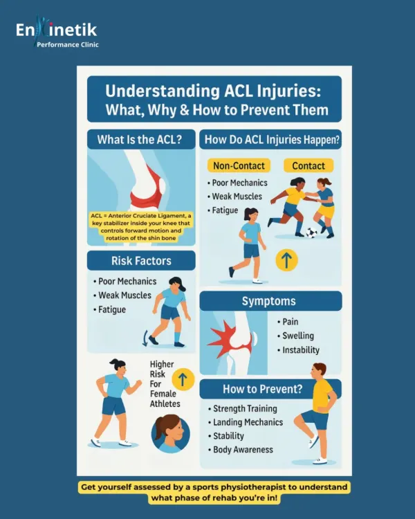 14
14Lower Body Sports Injury Treatment and Recovery
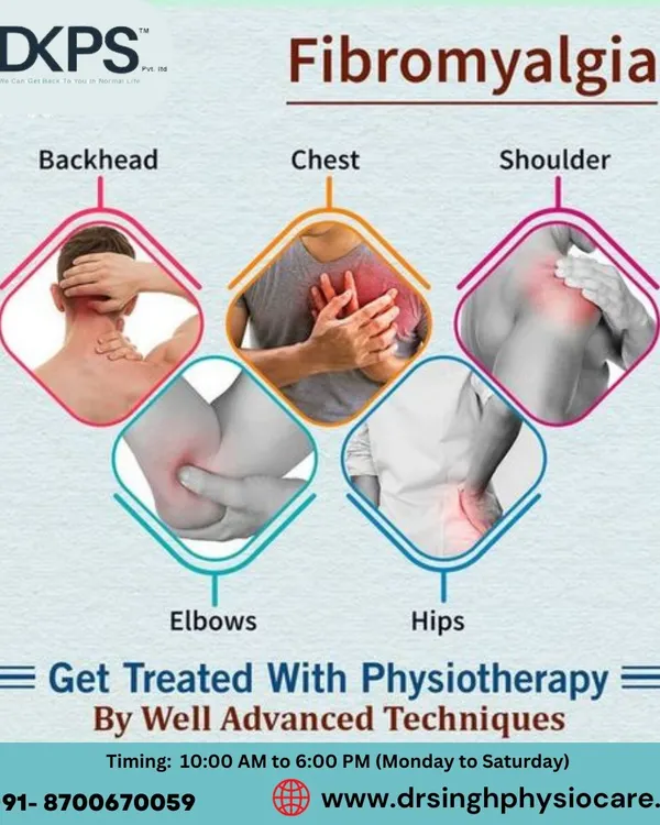 7
7
Understanding Pain and Our Healing Techniques
More from Sports Physiotherapy & Performance by AB Physiotherapy Clinic
More services by AB Physiotherapy Clinic